I. Skeletal System
A. Overview
1. Functions
2. Anatomy of a Long Bone
B. Bone Growth, Remodeling, and Repair
C. Bones of the Axial Skeleton
1. The Skull
2. The Hyoid Bone
3. The Vertebral Column
4. The Rib Cage
D. Bones of the Appendicular Skeleton
E. Articulations
II. Muscular System
A. Overview
1. Types of muscles
2. Functions
3. Skeletal muscles of the body
B. Skeletal muscle fiber contraction
1. Muscle fibers
2. Control of contraction
C. Whole muscle contraction
1. motor units
2. energy
3. fast twitch and slow twitch
D. Muscular Disorders
E. Homeostasis
Bones, cartilage, and fibrous connective tissue are all components of the skeletal system. The skeleton plays many important roles in the body. It supports the body, protects soft body parts, produces blood cells, stores minerals and fat, and permits flexible body movement in conjunction with the muscles.

The main portion of a long bone, or shaft, is called the diaphysis. Inside is a large medullary cavity, composed of compact bone. Compact bone is very highly organized and contains osteons, which are tubular units. Spongy bone is very unorganized in appearance. Cartilage is more flexible than bone, but not as strong. It is made up of cells called chondrocytes. There are three types of cartilage : hyaline, fibrocartilage, and elastic cartilage. Fibrous connective tissue contains cells called fibroblasts and makes up ligaments and tendons.
There are several different types of cells that are involved in growth, remodeling, and repair of bones. Osteoblasts are bone forming cells that promote sending calcium salts into the matrix. Osteocytes come from osteoblasts and maintain bone structure. Osteoclasts absorb bone cells. They break down bones.

The formation of a bone is called ossification. In a process called intramembranous ossification, bones develop in between the sheets of fibrous connective tissue. The most common type of ossification is endochondral ossification. During this process, bone replaces cartilaginous models of the bones gradually. Chondrocytes put down hyaline cartilage. As the cartilage model calcifies, the chondrocytes disappear. Osteocytes secrete bone matrix which undergoes calcification. This forms a bone collar, which covers the diaphysis. Blood vessels lay down spongy bone. The spongy bone is absorbed by osteoclasts and becomes the medullary cavity. A band of cartilage called a growth plate is in between the primary center and all secondary centers. Once the growth plates close, growth in bone length no longer occurs.
Hormones, which are chemical messengers, are involved in bone growth. Vitamin D is converted to a hormone that helps with the intestinal absorption of calcium. When calcium can't be absorbed, children can develop a condition called rickets. Growth hormone stimulates growth of the growth plates.
Bone renewal, also known as remodelling, is when bones break down and reform. Up to 18% of the bones in our body are recycled every year. Bone recycling helps the body regulate how much calcium is in the blood.
Bone repair happens after a bone breaks or fractures. There are four steps of repairal. First, blood escapes and forms a hematoma between the broken bones. Then, a fibrocartilaginous callus fills the space between the ends of broken bone. Next, a bony callus joins the bones together. Finally, bone remodelling takes place.
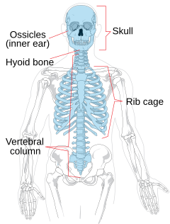
The axial skeleton is in the midline of the body. Its main components are the skull, the hyoid bone, the vertebral column, and the rib cage. The cranium and the facial bones make up the skull. The cranium is made up of eight bones that fit tightly together. It protects our brain. Some of the bones in the cranium contain the sinuses. The major bones of the cranium share the names of the lobes of the brain. The facial bones are what give our faces structure. The most noticeable of these are the mandible, the maxillae, the zygomatic, and the nasal bones. The hyoid bone is attached to the temporal bones and the larynx. It anchors the tongue and is where the muscles associated with swallowing attach. There are 33 vertebrae in the vertebral column. The vertebral column protects our spinal cord. In between the vertebrae are intervertebral disks, made up of fibrocartilage which serve the purpose of padding. The rib cage is also known as the thoracic cage and consists of the ribs and the sternum. It protects internal organs, yet is flexible. There are twelve pairs of ribs in the rib cage. The sternum protects the heart and the lungs.

The bones within the pectoral and pelvic girdles and their attached limbs make up the appendicular skeleton. There are two pectoral girdles in the body. Each has a scapula and a clavicle. These are the shoulder blade and the collarbone. The humerus comes off the scapula and the radius and ulna come off the humerus. The hand has many bones, causing it to be capable of great flexibility. The pelvic girdle is where our pelvis is. Our legs descend from this, containing the femur, and the tibia and fibula. The foot, like the hand, has many bones for flexibility.
Bones are jointed. These joints are either fibrous, cartilaginous, or synovial. Most fibrous joints are immovable. Cartilaginous joints are only slightly movable. Synovial joints permit free movement.

There are three types of muscle tissue in humans : smooth, cardiac, and skeletal. They all have cells called muscle fibers. Smooth muscle cells are spindle shaped and usually form sheets. Smooth muscle does not fatigue easily. Cardiac muscle is what goes around the heart and forms the heart wall.
Skeletal muscles have many functions. They support the body, make bones move, help maintain constant body temperature, assist movement in cardiovascular and lymphatic muscles, and help protect internal organs and stabilize joints. Skeletal muscles operate in pairs. The beginning of a muscle is on a stationary bone and the insertion is on a bone that moves.
muscles shorten when they contract.

Special names have been assigned to some of the components in a muscle fiber. The sarcolemma, sarcoplasm, and sarcoplasmic reticulum are the same as a plasma membrane, cytoplasm, and endoplasmic reticulum in any other cell. The sarcolemma forms T tubules that penetrate the cell and come into contact with parts of the sarcoplasmic reticulum. The sarcoplasmic reticulum encases thousands of myofibrils. Skeletal muscles have both light and dark bands. These bands are called striations. The thick bands are made up of myosin and the thin ones are made up of actin.
When the muscles are stimulated, calcium is released. The movement of actin filaments relating to myosin filaments is known as the sliding filament model. The entire region containing axons is known as the neuromuscular junction. There are two other types of proteins in an actin filament. These proteins are tropomyosin and troponin.

A nerve fiber combined with the muscle fibers that it innervates is a motor unit. All of the muscles in a motor unit contract at the same time. Maximal sustained contraction is called tetanus. It is desirable to have good muscle tone, with the muscle being firm and solid.
Muscles have four possible energy sources. These are muscle triglycerides, plasma fatty acids, blood glucose, and muscle glycogen. Muscle cells also store ATP which is needed for contraction. The CP pathway is the fastest way for muscles to produce ATP. Fermentation can produce two ATPs when glucose is broken down. Cellular respiration is another way that muscle cells can acquire more ATP.
Fast twitch muscle fibers are designed for strength and are usually anaerobic. Slow twitch fibers are steadier and more enduring. Delayed onset muscle soreness is muscles contracting while they are lengthening, and appears 24-48 hours after strenuous exercise.
Common muscular conditions are spasms, convulsions, cramps, facial tics, sprains, and strains. Tendinitis is when the gliding motion of a tendon is impaired and the tendon is inflamed. Bursitis is inflammation of a bursa (a sac filled with a smooth, slippery surface that helps muscles and tendons glide).
Muscular diseases can be more serious. Myalgia is chronic aching muscles. Muscular Dystrophy is characterized by progressive degeneration of muscles. Myasthenia gravis is an autoimmune disease affecting the muscles of the face, neck, and extremities. Lou Gehrig's disease, or ALS, is characterized by the gradual loss of certain abilities.
The skeletal and muscular systems work together to produce movement and to protect body parts. Bones store and release calcium and blood cells are produced in red bone marrow. Muscles help to maintain the temperature of the body by constricting or contracting.
I used images from the following sites
No comments:
Post a Comment